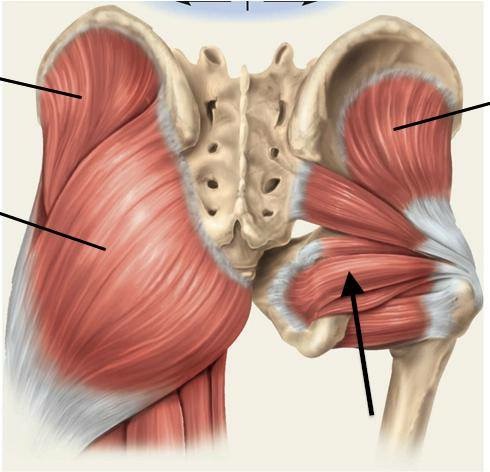
Left Hip Muscles Anatomy / Anatomy of the Left Hip and Leg - TrialExhibits Inc. - Back muscles of the hip.
Dapatkan link
Facebook
X
Pinterest
Email
Aplikasi Lainnya
Left Hip Muscles Anatomy / Anatomy of the Left Hip and Leg - TrialExhibits Inc. - Back muscles of the hip.. Knee assessment and hip mechanics learn how hip and pelvis mechanics can influence the knee powered by physiopedia start course. Learn about hip muscles human anatomy with free interactive flashcards. This anatomical atlas was especially designed for a specific public (radiologists, surgeons, rheumatologists and physicians specializing in musculoskeletal imaging). Your email address will not be published. The muscles of the neck can be divided into groups according to their location.
There are a lot of muscles of the hip and thigh. 3 months later i got acute excrutiating pain in inguinal area. The muscles of the neck can be divided into groups according to their location. The cavity of the acetabulum the external obturator muscle is short external rotator muscle of hip joint. Each muscle below has the bones in bold for intermediate learners and the specific bony landmarks for advanced learners.
The Deep Six, Turnout and Pilates from www.pilatesinstructoracademy.com The cavity of the acetabulum the external obturator muscle is short external rotator muscle of hip joint. Understanding the anatomy of the lower body, particularly the muscle locations and their functions, will help you to get the most from the exercises and programs presented on this website. Advanced hip flexor muscle anatomy. Rectus femoris muscle, one of the quadriceps muscles on the front of your thigh. Anterior muscles extend your legs and flex your thighs. In conclusion, a thorough understanding of pelvic and hip anatomy is important for. Attached to the bones of the skeletal system are about 700 named. It is a flat, triangular muscle on the anterior wall of the pelvis.
How muscles are named, 285 hints on how to deduce muscle actions, 286.
In order to isolate the abdominals, you need to minimize the involvement of the hip flexors and maximize the contraction of the abdominals. for detailed anatomy of pelvic bones, read anatomy of hip bone. Trunk muscles, 289 muscles of the thorax, 289 muscles of the abdominal wall, 289. The cavity of the acetabulum the external obturator muscle is short external rotator muscle of hip joint. Many doctors, no one believed there was anything wrong. Included within the chart are gorgeous illustrations of the pelvic diaphragm, sphincter muscles, gluteus maximus. Knee assessment and hip mechanics learn how hip and pelvis mechanics can influence the knee powered by physiopedia start course. The hip joint is the articulation of the pelvis with the femur, which connects the axial skeleton with the lower extremity. Rectus femoris forms the middle portion of the quadriceps. If left unchecked, this can lead to chronic knee pain from it band syndrome or acute yet severe injuries such as knee ligament tears (e.g. One example of an ab exercise that actually focuses. Rectus femoris muscle, one of the quadriceps muscles on the front of your thigh. The hip flexors are strong, powerful muscles that can overtake the abdominal muscles in some ab exercises.
Understanding the anatomy of the lower body, particularly the muscle locations and their functions, will help you to get the most from the exercises and programs presented on this website. Your email address will not be published. Major lower body muscle groups include leg and hip muscles, largest muscle groups in your body. There are a lot of muscles of the hip and thigh. I pulled some muscles on left hip hiking.
Muscles and Connective Tissues of the Hip and Upper Leg ... from classconnection.s3.amazonaws.com Trunk muscles, 289 muscles of the thorax, 289 muscles of the abdominal wall, 289. The cavity of the acetabulum the external obturator muscle is short external rotator muscle of hip joint. Attached to the bones of the skeletal system are about 700 named. Learn about hip muscles human anatomy with free interactive flashcards. If left unchecked, this can lead to chronic knee pain from it band syndrome or acute yet severe injuries such as knee ligament tears (e.g. Hip anatomy, function and common problems. In order to isolate the abdominals, you need to minimize the involvement of the hip flexors and maximize the contraction of the abdominals. Several muscles cross the front of the hip and create hip flexion, pulling the thigh and trunk toward each other, but probably the most important is the iliopsoas.
The cavity of the acetabulum the external obturator muscle is short external rotator muscle of hip joint.
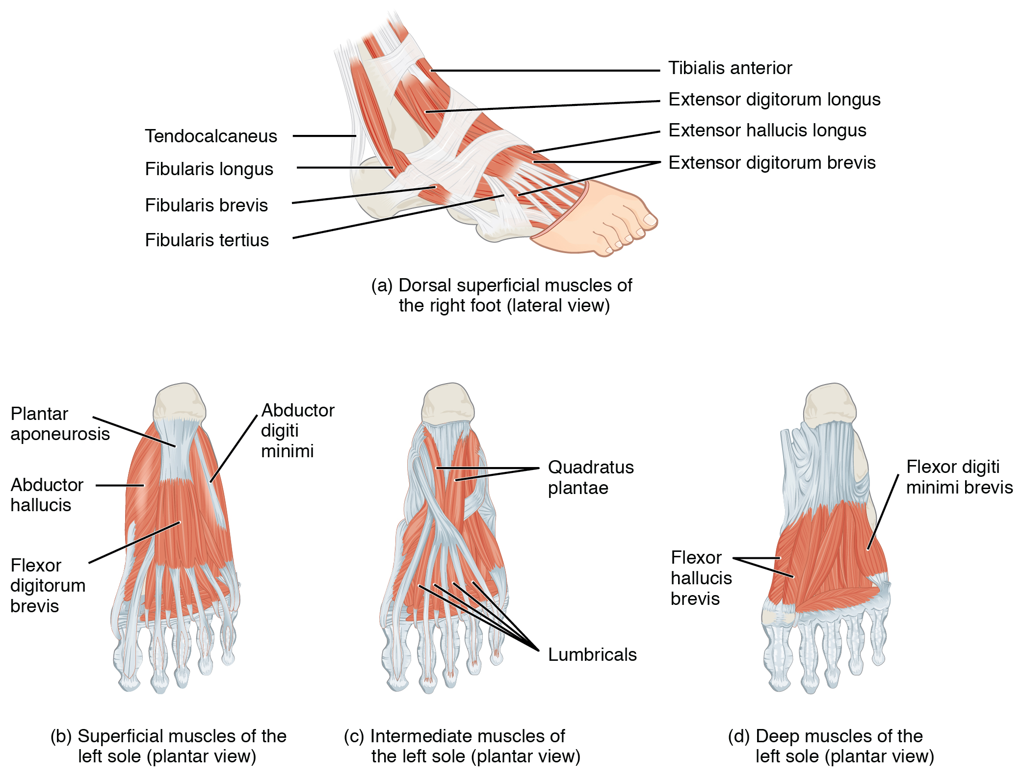
Microscopic anatomy of skeletal muscle. Understanding the anatomy of the lower body, particularly the muscle locations and their functions, will help you to get the most from the exercises and programs presented on this website. Muscles of the hips and thighs | human anatomy and. How muscles are named, 285 hints on how to deduce muscle actions, 286. The muscles of the pelvis, hip and buttock anatomical chart shows how each muscle in this area of the body works with the others, and the various minor systems within the major ones. If left unstretched, shortened hip flexors affect the position of the pelvis, which in turn affects the position and movement of the lower back. Attached to the bones of the skeletal system are about 700 named. Trunk muscles, 289 muscles of the thorax, 289 muscles of the abdominal wall, 289. Muscle movements, types, and names. The main functions of the neck muscles are to permit movements of the neck or head and to provide structural support of the head. Anatomy of the muscular system. If you know all the hip flexor names and bones they attach to, that's an awesome accomplishment! It is a flat, triangular muscle on the anterior wall of the pelvis.
Back muscles of the hip. Attached to the bones of the skeletal system are about 700 named. Many doctors, no one believed there was anything wrong. Advanced hip flexor muscle anatomy. Anatomy of a human body we study anatomy.
Appendicular Muscles of the Pelvic Girdle and Lower Limbs ... from philschatz.com In order to isolate the abdominals, you need to minimize the involvement of the hip flexors and maximize the contraction of the abdominals. Anatomy of a human body we study anatomy. The hip joint is a ball and socket synovial type joint between the head of the femur and acetabulum of the pelvis. Anterior muscles extend your legs and flex your thighs. I pulled some muscles on left hip hiking. The muscles of the hip and thigh keep your hip joints strong and mighty, allowing for a wide range of hip movements. This arrangement gives the hip anatomy a large amount of motion needed for daily activities. If left unstretched, shortened hip flexors affect the position of the pelvis, which in turn affects the position and movement of the lower back.
Learn their anatomy efficiently and easily using kenhub's muscle anatomy and reference charts!
Leave a reply cancel reply. Many doctors, no one believed there was anything wrong. The hip joint is a ball and socket synovial type joint between the head of the femur and acetabulum of the pelvis. Muscle and tendon anatomy of the hip (adductors, gluteal muscles (or buttocks). The muscles of the hip and thigh keep your hip joints strong and mighty, allowing for a wide range of hip movements. One example of an ab exercise that actually focuses. The main functions of the neck muscles are to permit movements of the neck or head and to provide structural support of the head. In order to isolate the abdominals, you need to minimize the involvement of the hip flexors and maximize the contraction of the abdominals. Human anatomy hip muscles anatomy anatomy study. Elbow muscles are commonly referred to as flexors or extensors, depending on how they affect elbow movement. This anatomical atlas was especially designed for a specific public (radiologists, surgeons, rheumatologists and physicians specializing in musculoskeletal imaging). The muscles of the pelvis, hip and buttock anatomical chart shows how each muscle in this area of the body works with the others, and the various minor systems within the major ones. These muscles constitute the anatomical classification known as the medial compartment of the thigh.
If you are looking for printable blank word search grid paper word search printable you've came to the right web. We have 100 Pics about printable blank word search grid paper word search printable like printable blank word search grid paper word search printable, printable blank crossword grid printable crossword puzzles and also spelling word word search grid by nats classroom tpt. Here you go: Printable Blank Word Search Grid Paper Word Search Printable Source: wordsearch-printable.com Play y ver 1,000 word searches, mazes, unscramble words, word games and others. We're celebrating the luck o' the irish with this fun st. Printable Blank Crossword Grid Printable Crossword Puzzles Source: crosswordpuzzles-printable.com Create fun, play when you want, hidden words in a word grid is fun. Have fun and print one or more of these great printable word search out today. Teacherfieracom Word Sear...
If you are looking for free printable alphabet individual alphabet letters you've came to the right page. We have 100 Pictures about free printable alphabet individual alphabet letters like free printable alphabet individual alphabet letters, individual alphabet letters clipart 10 free cliparts and also letter clipart printable letter printable transparent. Here you go: Free Printable Alphabet Individual Alphabet Letters Source: clipground.com Then start stapling the individual letters from the middle out so it's centered the way . So i made these alphabet free printable letters. Individual Alphabet Letters Clipart 10 Free Cliparts Source: clipground.com Letter cards, letter tiles, large outline letters, dot marker letters, alphabet charts. 26 alphabets in 18 themes. Artbyjean Paper Crafts Individual Letters And Numbers Source: 2.bp.blogspot.com Use these letters in...
One of the finest ways to find complimentary and high-quality free pharmacy technician math worksheets downloads is to start by searching online. The internet is home to a wide variety of websites that offer free free pharmacy technician math worksheets downloads, along with templates, coloring pages, and more. One methods to find these websites is to use a search engine, such as Google or Bing, and enter relevant keywords, such as "free free pharmacy technician math worksheets downloads" or "free free pharmacy technician math worksheets templates." This will educate a list of websites that offer free downloads, among other things blogs, online stores, and even government websites. Finding free download free pharmacy technician math worksheets can be easy and accessible, you can use the browser and visit websites that specialize in offering free resources. Be choosy about the websites you visit, choose trustworthy sites that offer high-quality, acc...


Komentar
Posting Komentar